
Welcome to the Anatomy and Physiology Lab Manual, your guide to exploring the human body through interactive lab exercises and hands-on activities. This manual provides a comprehensive overview of key biological systems, offering practical experiences to enhance your understanding of anatomical structures and physiological processes. Through detailed experiments and observations, you will gain essential skills in laboratory techniques and data analysis, fostering a deeper appreciation for the complexities of the human body. Get ready to engage in active learning and discover the wonders of anatomy and physiology firsthand!
1.1. Overview of the Lab Manual
This lab manual is designed to complement your anatomy and physiology coursework, offering a structured approach to hands-on learning; Organized into clear sections, it covers essential topics such as the skeletal, muscular, circulatory, and nervous systems, among others. Each chapter includes detailed lab exercises, step-by-step instructions, and interactive activities to reinforce theoretical concepts. The manual emphasizes practical application, encouraging students to explore anatomical structures and physiological processes in a controlled, educational environment. This resource is tailored to enhance understanding and retention through active participation and real-world examples.
1.2. Importance of Lab Work in Anatomy and Physiology
Lab work is essential for understanding anatomy and physiology, as it bridges theoretical knowledge with practical application. Hands-on activities allow students to observe and interact with biological structures, enhancing their comprehension of complex concepts. Through dissections, experiments, and data collection, students develop critical thinking and analytical skills. Lab experiences also foster teamwork and scientific inquiry, preparing learners for real-world applications in healthcare and research. Engaging in lab work creates a dynamic learning environment that strengthens retention and deepens understanding of human biology.

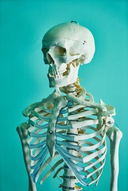
Skeletal System
The skeletal system provides structural support, facilitates movement, and protects vital organs. It consists of bones, cartilage, and ligaments, working together to maintain posture and enable bodily functions.
2.1. Bones and Joints: Structure and Function
Bones provide structural framework, protection, and leverage for movement. They are classified as long, short, flat, irregular, and sesamoid, each serving unique roles. Joints, or articulations, connect bones, enabling mobility. There are three types: synovial, cartilaginous, and fibrous joints. Synovial joints, like the knee, offer the greatest range of motion due to synovial fluid and cartilage. Bones and joints work together to support body weight, facilitate movement, and maintain posture, forming the foundation of the skeletal system’s functional capabilities.
2.2. Lab Exercises: Identifying Bones and Joints
3.1. Types of Muscles and Their Functions
Muscles are categorized into three types: skeletal, smooth, and cardiac. Skeletal muscles, attached to bones, enable voluntary movements and maintain posture. Smooth muscles, found in internal organs, function involuntarily, regulating processes like digestion and blood pressure. Cardiac muscle, exclusive to the heart, ensures continuous, rhythmic contractions for blood circulation. Each type differs in structure and control, but all contribute to movement, stability, and essential bodily functions, illustrating the diversity and complexity of the muscular system.

Muscular System
The muscular system is a vital body system responsible for movement, maintaining posture, and regulating body temperature through contractions of muscles. It works in conjunction with the skeletal system to enable voluntary and involuntary actions, essential for daily functions and overall mobility. This section explores its structure, function, and related lab exercises.
The muscular system comprises three types of muscles: skeletal, smooth, and cardiac. Skeletal muscles are voluntary, attached to bones, enabling movement and posture. Smooth muscles are involuntary, found in organs like the digestive tract, regulating processes like digestion. Cardiac muscle is specialized for the heart, ensuring continuous pumping of blood. Each type has distinct structures and functions, working together to facilitate movement, maintain organ function, and support overall bodily operations efficiently.
3.2. Lab Activities: Muscle Dissection and Identification
In this lab, students engage in muscle dissection to explore the structure and function of skeletal muscles. Using specimens or models, participants identify key muscle groups, such as flexors and extensors, and observe their attachments to bones; Activities include measuring muscle fibers, analyzing their arrangement, and documenting findings. This hands-on experience enhances understanding of muscle physiology and prepares students for clinical applications. Safety protocols and proper instrument handling are emphasized throughout the exercise.

Circulatory System
The circulatory system, comprising the heart, blood vessels, and blood, is essential for transporting oxygen, nutrients, and hormones while removing waste products. It maintains homeostasis and supports overall health by ensuring proper distribution of resources throughout the body. This system is vital for sustaining life and enabling cellular functions. Understanding its structure and function is crucial for grasping human physiology.
4.1. Blood Components and Blood Pressure
Blood consists of plasma, red blood cells (RBCs), white blood cells (WBCs), and platelets, each serving unique roles in oxygen transport, immunity, and clotting. Blood pressure, measured in millimeters of mercury (mmHg), reflects the force of blood against artery walls. Systolic pressure (top number) indicates pressure during heart contractions, while diastolic pressure (bottom number) measures relaxation. Factors like heart rate, vessel elasticity, and lifestyle influence blood pressure, which is crucial for maintaining proper circulation and overall health.
4.2. Lab Exercises: Blood Typing and Heart Rate
In this lab, students learn to determine blood type using anti-A and anti-B serums, observing agglutination reactions to identify ABO blood groups. This exercise emphasizes the importance of blood typing in medical procedures. Additionally, heart rate is measured to understand its relationship with physical activity and stress. Students record radial or carotid pulse rates at rest and after exercise, analyzing how lifestyle factors influence cardiovascular health. These hands-on activities provide practical insights into circulatory system functions and their real-world applications.

Respiratory System
The respiratory system facilitates gas exchange, enabling oxygen intake and carbon dioxide expulsion. Key organs include the nose, trachea, bronchi, and lungs. Breathing is essential for cellular respiration, sustaining life.
5.1. Structure and Function of the Respiratory System
The respiratory system consists of the nose, pharynx, larynx, trachea, bronchi, and lungs. Air enters through the nose, passes through the trachea, and reaches the bronchi, which branch into bronchioles. Alveoli, tiny air sacs in the lungs, facilitate gas exchange: oxygen diffuses into the blood, and carbon dioxide is expelled. The diaphragm contracts during inhalation, expanding the chest cavity. This system is vital for delivering oxygen to cells and removing carbon dioxide, maintaining cellular respiration and overall bodily function.
5.2. Lab Activities: Measuring Lung Capacity
In this lab, students measure lung capacity using spirometry to assess tidal volume, vital capacity, and residual volume. Participants inhale deeply and exhale into a spirometer to record air volumes. Additionally, a water displacement method involves breathing into a container filled with water to measure lung capacity. These activities help understand respiratory function and correlate lung volumes with anatomical structures. Safety precautions, such as proper equipment sterilization and student comfort, are emphasized to ensure accurate and reliable results.

Nervous System
The nervous system consists of the central and peripheral nervous systems, controlling voluntary and involuntary actions. It includes the brain, spinal cord, nerves, and sensory receptors, regulating body functions.
6.1. Basic Structure of the Nervous System
The nervous system is divided into the central nervous system (CNS) and peripheral nervous system (PNS). The CNS includes the brain and spinal cord, protected by the meninges and cerebrospinal fluid. The PNS consists of nerves connecting the CNS to the body. Neurons, the functional units, transmit signals through axons and dendrites. Neuroglial cells support neuronal function. The PNS is further divided into sensory (afferent) and motor (efferent) nerves, facilitating communication between the CNS and the body’s sensory receptors and effectors.
6.2. Lab Exercises: Reflex Testing and Neuron Structure
Lab exercises involve testing reflexes to understand nervous system function. Students observe and record reactions, such as patellar reflexes, to demonstrate neural pathways. Neuron structure is studied using microscope slides, identifying dendrites, cell bodies, and axons. Activities include labeling diagrams and simulating neural signaling. These exercises provide hands-on experience with reflex arcs and neuronal components, reinforcing concepts of neural communication and control. Practical application enhances understanding of the nervous system’s role in responding to stimuli and maintaining body functions.

Digestive System
The digestive system consists of organs like the mouth, esophagus, stomach, liver, pancreas, small intestine, and large intestine, responsible for breaking down food, absorbing nutrients, and eliminating waste.
7.1. Components of the Digestive System
The digestive system comprises the mouth, esophagus, stomach, small intestine, and large intestine. The mouth initiates digestion with teeth and enzymes, while the esophagus transports food to the stomach. The stomach churns food with gastric juices, breaking it down further. The small intestine absorbs nutrients into the bloodstream, and the large intestine absorbs water, forming waste. Accessory organs like the pancreas and liver provide digestive enzymes and bile, aiding in nutrient breakdown and absorption. Together, these components ensure efficient digestion and nutrient utilization.
7.2. Lab Activities: Enzyme Testing and Digestion Simulation
In this lab, students explore enzymatic action by testing amylase and pepsin activity using starch and protein substrates. Observing color changes with pH indicators demonstrates enzyme specificity. Additionally, a digestion simulation uses a plastic bag “stomach” filled with crackers, acid, and enzymes to mimic gastric digestion. Students record observations, linking enzyme function to real-world digestive processes. These hands-on activities clarify how enzymes break down nutrients and prepare them for absorption in the small intestine.
Key Concepts and Answers
This section summarizes key concepts, answers common questions, and provides tips for understanding lab experiments, helping students excel in their anatomy and physiology studies effectively.
8.1. Common Lab Questions and Answers
Here, we address frequently asked questions about lab exercises, such as identifying anatomical structures, using lab equipment, and interpreting results. Q: How do I identify bones accurately? A: Use diagrams and models for comparison. Q: Why is safety important in the lab? A: To prevent accidents and ensure a safe learning environment. These answers provide clarity and support for successful lab experiences.
- Understand the purpose of each exercise before starting.
- Record observations carefully for accurate data analysis.
8.2. Tips for Understanding Lab Experiments
To excel in lab experiments, start by thoroughly understanding the objectives and materials. Review pre-lab reading to familiarize yourself with procedures and expected outcomes. Ask questions during demonstrations to clarify doubts. Follow safety protocols to ensure a secure environment. Observe carefully and record accurate data to draw meaningful conclusions. Practice critical thinking to connect observations with theoretical concepts, enhancing your learning experience.
- Stay organized with labeled materials and equipment.
- Collaborate with peers to share insights and verify findings.
Additional Resources
Explore recommended textbooks, online tools, and interactive guides to enhance your anatomy and physiology learning experience. These resources provide comprehensive support for both study, experimentation, and practical application.
9.1. Recommended Textbooks and Online Tools
Enhance your learning with essential textbooks like Gray’s Anatomy and Anatomy: A Photographic Atlas. Online platforms such as Kenhub and AnatomyTOOL offer interactive 3D models and practice quizzes. Utilize digital resources like Visible Body and Labster for immersive simulations and virtual lab experiences. These tools complement your lab manual, providing visual and hands-on opportunities to deepen your understanding of anatomical structures and physiological processes. They are invaluable for reinforcing concepts and preparing for exams or lab activities.
9.2. Lab Safety Guidelines
Adhering to lab safety is crucial for a secure learning environment. Always wear protective gear like gloves and goggles when handling specimens or chemicals. Familiarize yourself with emergency procedures, such as the location of fire extinguishers and eye wash stations. Properly dispose of biological waste and clean workstations after use. Follow instructions carefully and avoid unauthorized equipment usage. Maintain a clutter-free workspace to prevent accidents. Report any hazards or incidents immediately to ensure a safe and efficient lab experience for everyone involved.
Completing this lab manual marks the end of an engaging journey through anatomy and physiology, providing a solid foundation for future studies and practical applications in the field.
10.1. Summary of Key Takeaways
This manual has guided you through a comprehensive exploration of the human body’s systems, emphasizing hands-on learning and practical application of anatomical and physiological concepts. Key takeaways include understanding the structure-function relationships in systems like skeletal, muscular, circulatory, respiratory, nervous, and digestive. Lab exercises such as bone identification, muscle dissection, blood typing, and lung capacity measurements have reinforced theoretical knowledge. By mastering these skills, you’ve gained a deeper appreciation for the intricate mechanisms that sustain life and maintain overall health.
10.2. Final Thoughts on Mastering Anatomy and Physiology
Mastering anatomy and physiology requires a blend of curiosity, dedication, and hands-on practice. This lab manual has provided a foundational understanding of the human body’s intricate systems and their interconnections. By engaging in lab exercises and reflective learning, you’ve cultivated essential skills in observation, analysis, and critical thinking. Remember that anatomy and physiology are dynamic fields, continually evolving with new discoveries. Stay curious, embrace lifelong learning, and apply these principles to advance your understanding of the remarkable human body.